

/ News, Research
Nanotags for more precise cancer surgery and diagnostics: Gold nanoparticles with optimized coatings to target ovarian cancer cells
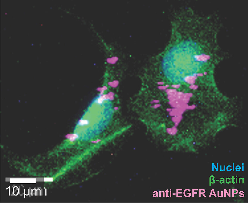
Researchers from the SNI Network have developed a straightforward surface-modification strategy for gold nanoparticles used in Surface-Enhanced Raman Scattering (SERS). The resulting nanoparticles, so called nanotags, can specifically recognize cancer cells and generate a measurable optical signal, offering potential for more precise cancer detection and future surgical applications. In a recently published study in the scientific journal Materials Today Bio, the researchers report a two-step coating strategy using two polyethylene glycol (PEG) conjugates carrying either a Raman reporter or an antibody. This carefully balanced coating enables the nanotags to recognize cancer cells expressing a specific biomarker while maintaining a stable SERS signal.
In SERS imaging, a laser is directed onto the nanotags after they have specifically bound to cancer cells. The gold nanoparticles strongly enhance the Raman signals of molecules located near their surface. This creates a characteristic optical fingerprint that can be detected with a Raman microscope and potentially be used to identify specific cells or tissues.
To ensure that the nanotags specifically target cancer cells, they are functionalized with antibodies that recognize structures on the cell surface. In this study, the researchers targeted the epidermal growth factor receptor (EGFR), a protein that is expressed on the surface of certain ovarian cancer cells. At the same time, the nanotags need to remain stable and avoid unwanted aggregation so that they retain their functionality.
Combining strong SERS signal enhancement, specific targeting and nanoparticle stability in a single nanotag is challenging. Researchers from the FHNW School of Life Sciences, the Department of Biomedicine at the University Hospital Basel and the Department of Chemistry at the University of Basel have now developed a two-step coating process for 60 nm gold nanoparticles that addresses these challenges.
Optimized surface coating
The teams led by Dr. Sina Saxer (FHNW), Dr. Francis Jacob (University Hospital Basel) and Prof. Dr. Jonathan de Roo (University of Basel) use an initial surfactant coating to temporarily stabilize the gold nanoparticles. This step allows the nanotags to remain stable in the buffer solutions required for attaching the anti-EGFR antibodies. A protective PEG coating is then added to complete the surface modification.
“Temporary stabilization ensures that the nanoparticles remain stable and prevents aggregation during the surface immobilization of the anti-EGFR antibodies,” explains Alessandro Stumpo, first author of the publication in Materials Today Bio and doctoral student at the SNI PhD School. “In our experiments, approximately three-quarters of the nanotags carried EGFR antibodies, while the binding sites remained active and accessible.”
The researchers demonstrated the specificity of the nanotags in in vitro experiments. The nanotags generated different SERS signals when applied to EGFR-positive and EGFR-negative ovarian cancer cells, demonstrating their ability to distinguish between cells based on the presence of the target receptor.
This optimized coating strategy provides a promising basis for developing SERS nanotags for more precise cancer diagnostics. In the future, such nanotags could potentially be used during surgery to help identify tumor tissue and distinguish it from surrounding healthy tissue.
Original publication:
Two-Step PEG-Based Surface Functionalization of Gold Nanoparticles Enables Specific SERS Imaging of EGFR in an Ovarian Cancer Cell Model
Alessandro Stumpo, Ricardo Coelho, Monica Nunez Lopéz, Lucy Kind, Jonathan De Roo, Francis Jacob, Sina Saxer
Materials Today Bio, 2026, 103602, https://doi.org/10.1016/j.mtbio.2026.103602