/ News
Tiny fish under a giant camera
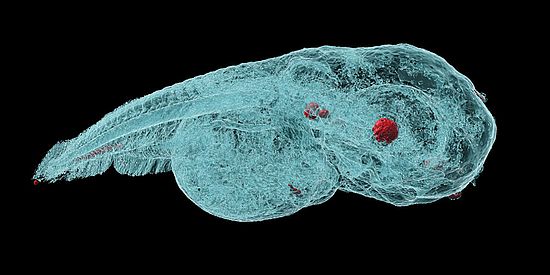
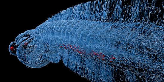
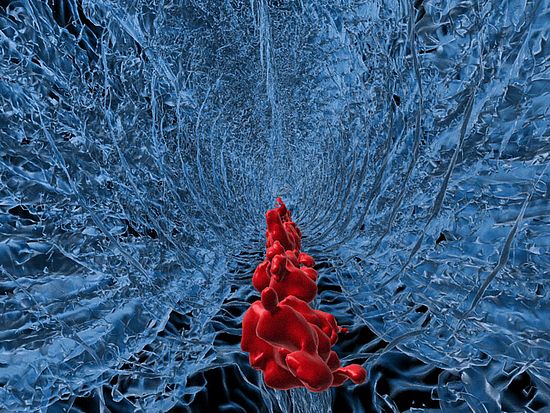
Using microtomography in phase contrast mode, researchers at the University of Basel succeeded in taking high-resolution, three-dimensional images of zebrafish embryos in which the distribution of nanoparticles (red) is visible. Also in red and easy to distinguish because of their high density are the lens of the eye and the otoliths in the inner ear – that is, tiny pieces of calcium carbonate in the vestibular system (Image: University of Basel, Jan Bolten)
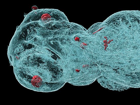
Head of a zebrafish embryo. The lens of the eye, otoliths and, on the right, an accumulation of the metal-based nanoparticles can be seen in red. (Image: University of Basel, Jan Bolten)


Metal-based nanoparticles are a promising tool in medicine – as a contrast agent, transporter of active substances, or to thermally kill tumor cells. Up to now, it has been hardly possible to study their distribution inside an organism. Researchers at the University of Basel have used a three-dimensional imaging method to take high-resolution captures inside zebrafish embryos.
To T establish the effectiveness and safety of metal-based nanoparticles for medical applications, developers must investigate the particle distribution inside a living organism and detect their accumulation. Until now, the particles have had to be labeled with radioactive or fluorescent markers. Such marker molecules, however, can influence the distribution of the nanoparticles in the organism. In addition, a high dose of particles is needed and distorts the results.
Researchers working with Professor Jörg Huwyler and Professor Bert Müller of the University of Basel recently reported an approach, that circumvents these problems, in the journal Small: they used microtomography in phase contrast mode based on synchrotron X-ray radiation to study the distribution of nanoparticles – without any special labeling – in zebrafish embryos.
The researchers used the synchrotron light source at the Paul Scherrer Institute to capture the images. “This is the biggest camera in Switzerland for these tiny creatures,” Huwyler says. “The phase contrast makes the individual organs and even the optic nerve visible,” Müller adds.
The magnetic iron oxide nanoparticles used in the study throw tissue differences in magnetic resonance tomography into sharper relief. In future, they could also be used to transport active substances to the desired site in the body directed by external magnetic fields.
Giant camera, 138 meters in diameter
Zebrafish embryos are particularly suitable for toxicology studies because their bodies are transparent and they have an immune system similar to that of humans. Even though the animals are barely three millimeters long, every cell in their bodies and the distribution of the injected nanoparticles can be discerned in the three-dimensional images.
The nanoparticles consist of a coated metal core. The coatings can be adapted to the desired application. The presented method makes it easier to investigate how particles with a particular coating will behave in an organism.
Original source
Emre Cörek, Griffin Rodgers, Stefan Siegrist, Tomaz Einfalt, Pascal Detampel, Christian M. Schlepütz, Sandro Sieber, Pascal Fluder, Georg Schulz, Harald Unterweger, Christoph Alexiou, Bert Müller, Maxim Puchkov, and Jörg Huwyler
Shedding Light on Metal-Based Nanoparticles in Zebrafish by Computed Tomography with Micrometer Resolution
Small (2020), doi: 10.1002/smll.202000746
Additional information and images can be found here
Further information
Prof. Dr. Jörg Huwyler, University of Basel, Department of Pharmaceutical Sciences, phone +41 61 207 15 13, email: joerg.huwyler@clutterunibas.ch
Prof. Dr. Bert Müller, University of Basel, Biomaterials Science Center, phone +41 61 207 54 30, email: bert.mueller@clutterunibas.ch