/ Erfolgsgeschichten
Rapid determination of three-dimensional structures – Nano Argovia project A3EDPI meets with a positive response
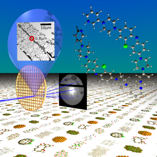
On a grating for transmission electron microscopy (TEM), a tiny crystal of the methylene blue derivative is placed. The tiny crystals are irradiated with electrons (blue beam) and a typical diffraction pattern is created. Based on the specific information of the diffraction pattern, the scientists can determine the chemical structure of the molecule. Numerous structures are waiting to be identified using this new method (Image: Tim Grüne).


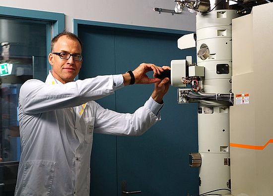
For over twelve years, the Swiss Nanoscience Institute (SNI) has supported applied research projects as part of the Nano Argovia program, providing companies in Northwestern Switzerland with access to innovative research through the SNI network. As these projects focus on applications that are useful to the companies, they rarely lead to publications in prominent scientific journals. The Nano Argovia project A3EDPI, led by Dr. Tim Grüne of the Paul Scherrer Institute (PSI), is an exception. Together with colleagues, Grüne recently published some initial results in the journal Angewandte Chemie, and their article triggered a huge response. In it, the scientists described how they successfully use electron nanocrystallography to determine the three-dimensional structure of active pharmaceutical ingredients in powder form.
Powder is hard to analyze
To develop new active pharmaceutical ingredients efficiently and to obtain a license for their use, researchers need to know the exact three-dimensional structures of the substances, since the efficacy of a compound depends on its spatial configuration. If the active substances exist as individual crystals, the 3D structure can be determined using X-ray structure analysis. In many cases, however, the scientists have to work with powders – in other words, mixtures of crystalline nanograins measuring just 100–500 nanometers across. Until now, reliably analyzing their 3D structure required a great deal of time and instrumentation.
Electron beams reveal three-dimensional structure
Now, as part of the Argovia project A3EDPI, an interdisciplinary team of scientists from the Paul Scherrer Institute (PSI), the University of Basel, and ETH Zurich has collaborated with the companies Dectris AG and Crystallise! AG to investigate whether electron nanocrystallography is suitable for determining the 3D structure.
“We expose the samples to a high-energy beam of electrons,” explains project leader Dr. Tim Grüne (PSI). “As the electrons have wave properties, each molecule produces a highly specific diffraction pattern depending on the arrangement of the atoms, and this allows us to draw precise conclusions regarding the atomic structure.”
To begin with, the scientists developed a prototype of an electron diffractometer. Here, they combined an EIGER hybrid pixel detector from Dectris with a transmission electron microscope (TEM). As a test substance, they then analyzed the cold medicine Grippostad©, which contains a mixture of crystalline and non-crystalline, active and inactive components. “The small size of the crystals in this powder would prevent X-ray analysis from delivering satisfactory results,” explains Tim Grüne. “Using electron diffraction, however, we were able to identify the active substance unambiguously as acetaminophen, also known as paracetamol.”
Electron beam diffraction can also successfully determine the structure of larger and more complex chemical compounds, as the researchers have demonstrated using a new, unknown derivative of methylene blue.
Tim Grüne, who is currently working as a senior scientist at the PSI, is certain that the method will soon enjoy broad applications. In an interview with the journal Science, he says: “Pharmaceutical companies build up huge collections of crystalline compounds for their drug screening activities, but only around a quarter to a third of these compounds form crystals that can be analyzed using X-ray crystallography. Our method may help to bypass this bottleneck in the analysis and identification of new active substance candidates, as we can analyze even the very small crystals – just a few hundred nanometers in size – for which no 3D structure currently exists.”
Tim Grüne’s phone is currently ringing round the clock, as scientific journals such as Science and Nature are keen to learn more about the method. For example, Nature recently published an article entitled “Why didn’t we think to do this earlier? Chemists thrilled by speedy atomic structures,” and the journal Science also believes that the method could significantly speed up the development of new drugs as well as forensic investigations (see links).
The Nano Argovia project A3EDPI will conclude at the end of 2018. However, Tim Grüne will continue to use electron-beam diffraction to determine structures – albeit not at the PSI. At the start of next year, he will take over responsibility for X-ray structure analysis at the University of Vienna’s Faculty of Chemistry. Once there, he will hopefully continue his work on this successful approach, whose wide range of applications has now been demonstrated by the Nano Argovia project.
Original paper:
https://onlinelibrary.wiley.com/doi/abs/10.1002/anie.201811318
More information about the SNI’s Nano Argovia program:
https://nanoscience.ch/de/forschung/angewandte-forschung/