
SEM and Cryo-SEM – Scanning electron microscopy
We use SEM to examine the surface of a sample with secondary electrons (SE) and create a topographic image. We use backscattered electrons (BSE) to obtain information about qualitative material composition. Cryo-SEM can be used to image hydrated samples without drying artifacts by means of flash-freezing, or samples are dried beforehand by the critical point drying (CPD) procedure.

TEM – Transmission Electron Microscopy
In TEM, a specimen is irradiated with electrons and the electrons that emerge are projected onto a surface to form an image. Samples must be thin enough to ensure optimal irradiation. This process provides an insight into what is going on inside a specimen. Atomic resolution level can be achieved with STEM (scanning transmission electron microscopy).
AFM – Atomic Force Microscopy
With a tip attached to the end of a fine measuring beam, the surface is scanned on a micro- to nanometer scale. The forces that act between the sample surface and the tip can also be used to determine surface properties, such as adhesion, charge distribution, elasticity, magnetic or electrostatic field strength or surface conductivity.

LSM – 3D-Laser Scanning Microscopy
The color 3D laser scanning microscope determines surface data by point-by-point laser and white light scanning with an extremely large depth of field and high precision. Furthermore, profile, volume and roughness analyzes, as well as the thickness of a transparent objects can be determined.

Devices for light and fluorescence laser microscopy
On request, we also offer simple measurements with a confocal fluorescence laser microscope. This service is provided by us in the Imaging Core Facility of the Biozentrum.
For independent use of light and confocal fluorescence laser microscopes, please contact the Imaging Core Facility.

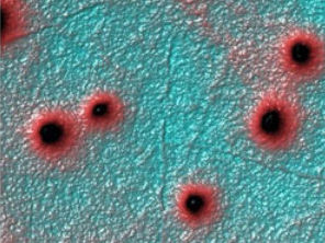
Digital photomontage and coloration
With the help of photo editing programs, we create collages from microscopic images or color your scanning electron microscopy images as desired. Please place a nonbinding inquiry here.
